Successful airway management depends on the ability to align the oral, pharyngeal, and laryngeal axes to maintain airway patency, whether through intubation or a less invasive approach. Neck mobility plays a central role in achieving this alignment, and any limitation can complicate intubation or increase the risk of failed airway attempts. For anesthesiologists and emergency providers, understanding the medical conditions that reduce cervical mobility is essential for planning safe airway strategies.
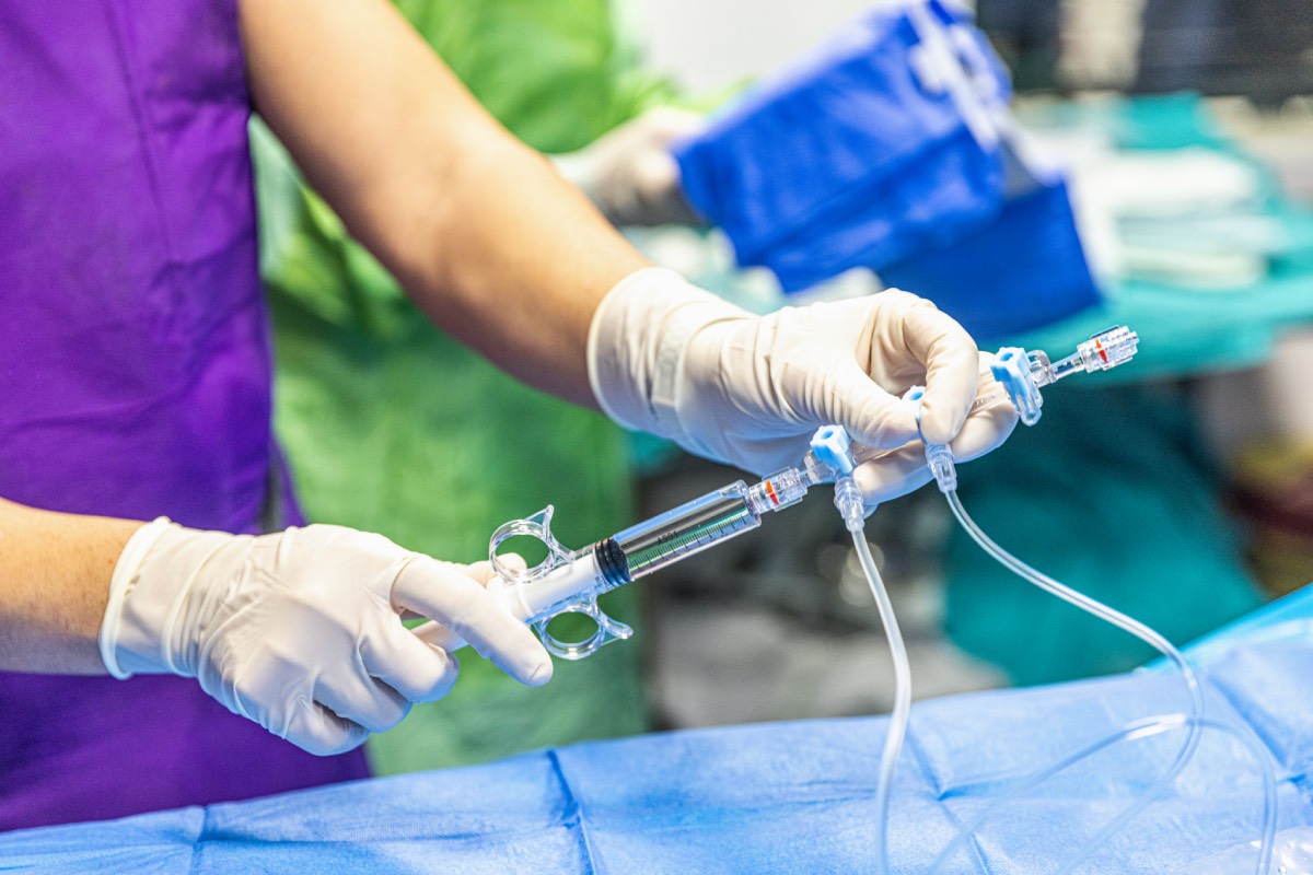
To obtain the best view of the vocal cords, the head and neck are usually positioned with the lower neck bent forward and the head tilted back, or the “sniffing position.” If a patient cannot move their neck to reach this position, it can make placing a breathing tube with a traditional laryngoscope more difficult. In this case, physicians may need to use other methods, like a video scope, a fiberoptic camera, or placing the tube while the patient is still awake 1–4.
Acute trauma is a common scenario in which neck mobility must be restricted. Patients with suspected cervical spine injury are managed with rigid immobilization to prevent spinal cord damage, and airway management must be performed without manipulating the neck, often using video laryngoscopy, fiberoptic scopes, or supraglottic devices as a bridge. In these circumstances, an additional challenge is avoiding incurring secondary neurologic injury 4–7.
Rheumatoid arthritis is a well-recognized cause of reduced neck mobility and can complicate airway management. Chronic inflammation leads to joint destruction, atlantoaxial instability, and limited extension at the atlanto-occipital joint. Osteoarthritis and ankylosing spondylitis can also restrict mobility by causing progressive stiffening and fusion of the cervical spine. In ankylosing spondylitis, the “bamboo spine” deformity results in a rigid column that cannot be manipulated, significantly complicating airway access. These patients often require awake fiberoptic intubation as the safest approach 8–11.
Certain congenital syndromes are associated with reduced cervical spine flexibility. For example, Klippel-Feil syndrome, characterized by congenital fusion of cervical vertebrae, results in short neck stature and markedly decreased motion. Additionally, patients with Down syndrome may have atlantoaxial instability, which necessitates extreme caution during neck extension. Awareness of these conditions is critical during preoperative evaluation, since their presence may not always be obvious without a thorough history or physical exam 12–15.
Patients who have undergone cervical spine surgery, such as fusion or instrumentation, often have restricted neck motion afterward. Similarly, individuals with head and neck cancers who have received radiation therapy may develop fibrosis and contractures that stiffen soft tissues and limit extension. In these cases, both bony and soft tissue restrictions contribute to difficult airway scenarios, and advanced planning is essential 16,17.
When neck mobility is impaired, conventional laryngoscopy is less reliable. Preoperative airway assessment should include an assessment of cervical range of motion along with standard predictors such as the Mallampati score and thyromental distance. Anticipating a difficult airway enables providers to prepare alternative devices, enlist experienced personnel, and consider awake or fiberoptic techniques. In emergencies, maintaining oxygenation with supraglottic devices may be life-saving when intubation proves impossible 18–21.
Conditions that decrease neck mobility, ranging from acute trauma to chronic rheumatologic, congenital, or postsurgical changes, pose significant challenges to airway management. Recognizing these limitations before induction of anesthesia or emergency intervention allows clinicians to select safer and more effective strategies. Ultimately, careful evaluation and preparation remain the cornerstone of successful airway management in patients with reduced cervical mobility.
References
1. Optimal Patient Positioning for intubation and airway management – The Anaesthesia Collective. https://www.anaesthesiacollective.com/optimal-patient-positioning-for-intubation-and-airway-management/ (2023).
2. Cook, T. M. & Chrimes, N. ‘Flextension’: a new term to describe optimal head and neck positioning for airway management. Anaesthesia 80, 220–221 (2025). DOI: 10.1111/anae.16484
3. Ezri, T. & Dukhan, A. Bedside predictors of difficult airway – neck mobility. Anaesthesia 74, 1616–1616 (2019). DOI: 10.1111/anae.14831
4. Aleksandrowicz, D. & Gaszyński, T. Airway Management with Cervical Spine Immobilisation: A Comparison between the Macintosh Laryngoscope, Truview Evo2, and Totaltrack VLM Used by Novices—A Manikin Study. Biomed Res Int 2016, 1297527 (2016). DOI: 10.1155/2016/1297527
5. Waseem, M., Torlincasi, A. M. & Hall, W. A. Cervical Injury. in StatPearls (StatPearls Publishing, Treasure Island (FL), 2025).
6. Austin, N., Krishnamoorthy, V. & Dagal, A. Airway management in cervical spine injury. Int J Crit Illn Inj Sci 4, 50–56 (2014). DOI: 10.4103/2229-5151.128013
7. Melesse, D. Y., Tesema, T. T., Mekonnen, Z. A. & Chekol, W. B. Airway management for individuals with suspected or confirmed traumatic cervical spine injuries: A comprehensive review and analysis. Perioperative Care and Operating Room Management 35, 100390 (2024). DOI: 10.1016/j.pcorm.2024.100390
8. Spinal Arthritis (Arthritis in the Back or Neck). https://www.hopkinsmedicine.org/health/conditions-and-diseases/spinal-arthritis (2024).
9. Cervical Spondylosis (Arthritis of the Neck) – OrthoInfo – AAOS. https://www.orthoinfo.org/en/diseases–conditions/cervical-spondylosis-arthritis-of-the-neck/.
10. What Is Ankylosing Spondylitis? Cleveland Clinic https://my.clevelandclinic.org/health/diseases/ankylosing-spondylitis.
11. Atlantoaxial Osteoarthritis. Physiopedia https://www.physio-pedia.com/Atlantoaxial_Osteoarthritis.
12. Ali, F. E., Al-Bustan, M. A., Al-Busairi, W. A., Al-Mulla, F. A. & Esbaita, E. Y. Cervical spine abnormalities associated with Down syndrome. Int Orthop 30, 284–289 (2006). DOI: 10.1007/s00264-005-0070-y
13. Menger, R. P., Rayi, A. & Notarianni, C. Klippel Feil Syndrome. in StatPearls (StatPearls Publishing, Treasure Island (FL), 2025).
14. Stupar, N. V. et al. Multiple Major and Minor Anomalies Associated With Klippel-Feil Syndrome: A Case Report. Arch Rheumatol 31, 082–086 (2016). DOI: 10.5606/ArchRheumatol.2016.5714
15. Foley, C. & Killeen, O. G. Musculoskeletal anomalies in children with Down syndrome: an observational study. Arch Dis Child 104, 482–487 (2019). DOI: 10.1136/archdischild-2018-315751
16. Koda, M. et al. Factors Significantly Associated with Postoperative Neck Pain Deterioration after Surgery for Cervical Ossification of the Posterior Longitudinal Ligament: Study of a Cohort Using a Prospective Registry. J Clin Med 10, 5026 (2021). DOI: 10.3390/jcm10215026
17. Jimenez-Socha, M. et al. Radiation-Induced Fibrosis in Head and Neck Cancer: Challenges and Future Therapeutic Strategies for Vocal Fold Treatments. Cancers (Basel) 17, 1108 (2025). DOI: 10.3390/cancers17071108
18. Jackson, J. S. & Rondeau, B. Mallampati Score. in StatPearls (StatPearls Publishing, Treasure Island (FL), 2025).
19. Thyromental Distance – an overview | ScienceDirect Topics. https://www.sciencedirect.com/topics/medicine-and-dentistry/thyromental-distance.
20. Zhang, K., Zhou, M., Zou, Z., Zhu, C. & Jiang, R. Supraglottic airway devices: a powerful strategy in airway management. Am J Cancer Res 14, 16–32 (2024). DOI: 10.62347/KJRU4855
21. Li, Q., Xie, P., Zha, B., Wu, Z. & Wei, H. Supraglottic jet oxygenation and ventilation saved a patient with ‘cannot intubate and cannot ventilate’ emergency difficult airway. J Anesth 31, 144–147 (2017). DOI: 10.1007/s00540-016-2279-x